|
Findings
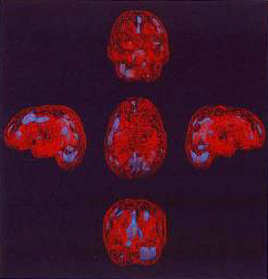
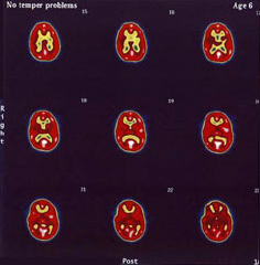
We found no temporal hypoperfusion in the 3-D images with only one cut-off or threshold of 60-65% of the maximum value in children with or without temper problems (Figure 1). Nor did we find any temporal hyperperfusion in the dual threshold 3D images in children without temper problems, although areas of increased flow could often be seen in the prefrontal, and posterior parietal cortex as well as in the cingulate gyrus (Figure 2 A). The location of the increased perfusion was more difficult to ascertain on the transverse views (Figure 2 B).
|
|
|
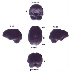
Figure 1. Volume rendered 3D surface images of a child with temper problems. No cortical temporal lobe change is appreciated. There is a non-specific cortical deficit in the vertex in the region of the paracentral lobules adjacent to midline.
|
|
|
|
|
Figure 2A
|
Figure 2B
|
|
Figure 2 A. Dual level 3D images. The red lattice work represents normal cortex that has perfusion of between 65-87% of maximum. The blue areas represent regions of highest perfusion ranging between 88-100% of maximum pixel counts. The vertex view (center image) shows high blood flow in the left frontal parietal region as well as in the anterior and posterior cingulate gyrus. The anterior view (top image) shows the increased blood flow in the anterior cingulate gyrus as well as the left superior pre-frontal region. The lateral images show the separation of the regions of high flow in the pre-frontal area from the anterior cingulate gyrus. The anterior (top) and posterior (bottom) views show the right cerebellar hemisphere has higher blood flow than the left. The temporal lobe blood flow is not increased. The transverse sections, B, show areas of highest flow as white with the transition between red and yellow at 60% of maximum roughly corresponding to gray and white matter demarcation. Increased flow can be seen in the region of the left insula (section number 20), the anterior and posterior cingulate, the left dorsal fronto-parietal region as well as the midline prefrontal area, the right parietal area and visual cortex. This child had no temper problems.
|
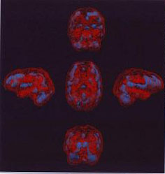
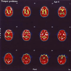
By contrast, in the dual threshold 3-D images with the upper cut-off of 88% and the lower cut-off of 60-65%, we observed an increase in the perfusion or activation in the region of the superior gyrus of either one or both temporal lobes (Figure 3A). It was also often striking on the transverse views as well (Figure 3B). This was detected in all of the eight patients with temper problems. It did not matter what clinical diagnosis they received from the referring physician. Increased perfusion in the temporal lobe was not seen in 10 patients with the same clinical diagnoses, but who did not have temper problems reported.
|
|
|
|
Figure 3A
|
Figure 3B
|
|
Figure 3A. Marked increased in blood flow can be seen in the dual level 3D images beginning in the dorsal pre-frontal cortex, affecting the superior temporal gyri bilaterally as well as the anterior and posterior portions of the cingulate gyrus. Activity in the cerebellar hemispheres is symmetric. The basal ganglia and thalami can be readily seen on the anterior (top) and vertex (center) views. The transverse views, B, show the increased blood flow (white is the highest value) ringing the cortex and affecting the temporal lobes in section 19. This child had temper problems.
|
|
|